ABC Imagem Cardiovasc. 2026; 39(3): e20260107
Increased Blood Pressure and Increased Optic Nerve Sheath Diameter: Intracranial Involvement or a Nonspecific Marker?

DOI: 10.36660/abcimg.20260107i
This Short Editorial is referred by the Research article "Ultrasound Measurement of Optic Nerve Sheath Diameter in Acute Hypertensive States For Detection of Intracranial Involvement: A Meta-Analysis".
Effects of increased blood pressure extend beyond the systemic arteries
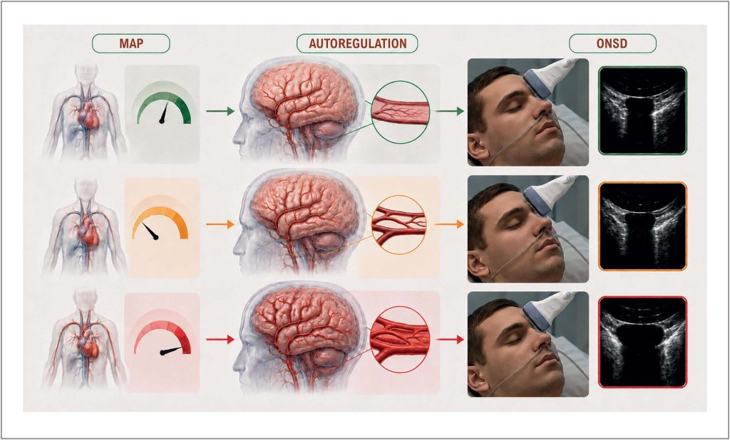
A hypertensive emergency is defined by presence of acute target-organ damage, not merely by an increased blood pressure reading. In brain, however, identifying such involvement at bedside remains challenging. Headache, nausea, visual disturbances, seizures, and altered consciousness are clinically important but nonspecific manifestations. Although fundoscopy has traditionally played an important role in clinical assessment, its reliability depends on adequate training, appropriate technical conditions, and patient cooperation. Moreover, papilledema may be absent during initial hours of increased intracranial pressure (ICP). Within this diagnostic window, ultrasonographic measurement of optic nerve sheath diameter (ONSD) represents an appealing option: it is rapid, portable, and repeatable and capitalizes on anatomical continuity between intracranial subarachnoid space and perioptic compartment.,
Article accompanying this short editorial provides most focused synthesis to date of ONSD findings in acute hypertensive states. Its results are consistent with those of previous studies involving acute hypertension, severe preeclampsia, and eclampsia.– Review included 14 studies comprising 995 participants, predominantly patients with preeclampsia or eclampsia, together with a considerably smaller group of nonpregnant patients with hypertension. Across nine studies with continuous data suitable for pooling comprising 426 hypertensive patients and 283 controls, ONSD was significantly greater in hypertensive group, with a large standardized mean difference (Hedges’g: 1.55; 95% CI: 1.06-2.04; p < 0.001). This is not a trivial finding: it suggests that association between acute hypertension and optic nerve sheath distension is sufficiently consistent that it cannot readily be dismissed as an isolated artifact.
[…]
Keywords: Arterial Pressure; Intracranial Pressure; Optic Nerve; Ultrasonography
15