ABC Imagem Cardiovasc. 2026; 39(3): e20260095
Cardiac Involvement in Fabry Disease: From Electrocardiography to Recent Advances in Cardiovascular Imaging

Abstract
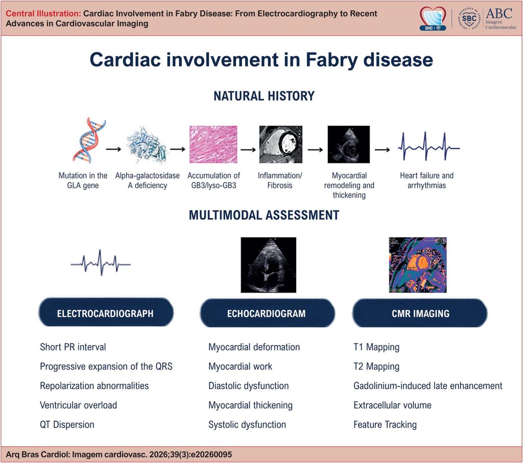
Fabry disease is an X-linked lysosomal storage disorder caused by α-galactosidase A deficiency, leading to the progressive accumulation of globotriaosylceramide (Gb3) and globotriaosylsphingosine (lyso-Gb3) in multiple organs. Cardiac involvement is the leading cause of morbidity and mortality, making early recognition of cardiomyopathy fundamental to optimizing treatment and improving prognosis. This review summarizes the current evidence on the role of electrocardiography, conventional and advanced echocardiography, and cardiac magnetic resonance (CMR) imaging in the assessment of cardiac involvement in Fabry disease. The main electrocardiographic findings, the structural and functional changes identified by echocardiography, including myocardial deformation by speckle tracking and myocardial work, as well as the contributions of magnetic resonance imaging to tissue characterization through late gadolinium enhancement (LGE), T1 and T2 mapping, extracellular volume (ECV), and feature tracking are discussed. The applications of these methods in the early detection of cardiomyopathy, differential diagnosis, risk stratification, monitoring of disease progression, and evaluation of treatment response are discussed. The integration of different imaging methods allows for a more comprehensive characterization of the pathophysiological mechanisms and stages of Fabry cardiomyopathy, contributing to early diagnosis, clinical decision-making, and longitudinal follow-up of these patients.
16