ABC Imagem Cardiovasc. 2026; 39(3): e20260069
Beyond the Appendage: Left Atrial Wall Thrombosis in Rheumatic Mitral Stenosis

DOI: 10.36660/abcimg.20260069i
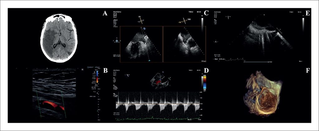
A 61-year-old woman with a history of rheumatic valvular heart disease and permanent atrial fibrillation (AF), chronically anticoagulated with rivaroxaban, presented with acute ischemic stroke and bilateral upper-limb ischemia. Neuroimaging revealed an acute infarction in the right middle cerebral artery territory and a subacute infarction in the left posterior cerebral artery territory (). Doppler ultrasound confirmed arterial occlusion in both upper limbs ().
Transesophageal echocardiography demonstrated marked left atrial (LA) enlargement, dense spontaneous echocardiographic contrast within the left atrial appendage (LAA), and reduced appendage flow velocities, without evidence of an organized thrombus (). The mitral valve exhibited features consistent with chronic rheumatic disease, with an estimated valve area of 1.0 cm² (). Notably, two- and three-dimensional imaging identified a large mural thrombus adherent to the LA wall, extending along the lateral and posterior walls and measuring approximately 4 × 0.8 cm (; Supplementary Videos 1-3).
[…]
Keywords: Atrial fibrillation; Echocardiography; Mitral valve stenosis
260