Arq Bras Cardiol: Imagem cardiovasc. 2021; 34(2): eabc172
Cardioverter-Defibrillator Implantation Through Persistent Left Superior Vena Cava
DOI: 10.47593/2675-312X/20213402eabc172
Case
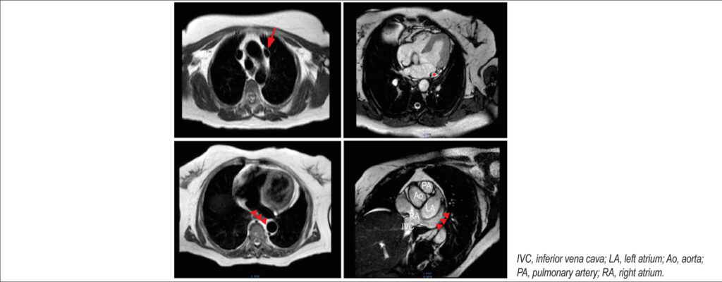
A 72-year-old woman with hypertension was admitted for sudden cardiac arrest secondary to idiopathic ventricular fibrillation. Cardiac magnetic resonance disclosed a persistent left superior vena cava (PLSVC) without other cardiovascular abnormalities for which an implantable cardioverter-defibrillator (ICD) was proposed with left access. Intraoperatively, cephalic vein cannulation placed the wire in the PLSVC that drained in the coronary sinus and subsequently the right atrium. A wide loop maneuver placed the lead tip facing the tricuspid valve, and right ventricle access was obtained with successful positioning of the ventricular lead. Device parameters were checked and considered suitable, and the procedure was finalized with active fixation, a fluoroscopy time of 1.35 minutes, and a radiation dose of 143.12 μGy/cm2. Normal pacing parameters were present at the 3-year follow-up.
PLSVC is a congenital venous anomaly present in 0.5% of the general population that is usually asymptomatic and incidental on invasive procedures or imaging. Although not a contraindication to ICD, lead placement is challenging because it has to negotiate two bends, one at the coronary sinus and the other in the tricuspid valve. Stylet shaping or wide loop techniques are reliable and present good outcomes not affecting pacing parameters on long-term follow-up.
[…]
134