ABC Imagem Cardiovasc. 2026; 39(2): e20260046
Mitral Valve Leaflet Hypoplasia in Adults: Role of Cardiovascular Imaging
DOI: 10.36660/abcimg.20260046i
Abstract
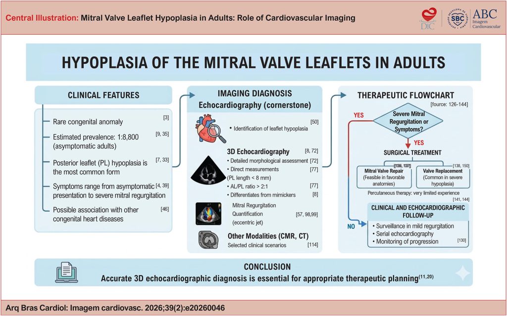
Mitral valve leaflet hypoplasia is a rare congenital anomaly, traditionally described in childhood but increasingly recognized in adults, often as an incidental finding or during the evaluation of mitral regurgitation. Clinical presentation is heterogeneous and depends on leaflet anatomy, the subvalvular apparatus, and the severity of regurgitation. In this narrative literature review, including case reports, case series, and review articles from nationally and internationally recognized journals, epidemiological and clinical aspects are discussed, with particular emphasis on echocardiographic findings. Posterior leaflet hypoplasia is the most common form and may be partial or complete. Three-dimensional echocardiography plays a central role in anatomical assessment, enabling direct measurements of leaflet area and length and helping differentiate true hypoplasia from mimicking entities such as mitral cleft, functional restriction, or subvalvular abnormalities. The estimated prevalence in asymptomatic adults is approximately 1:8,800. Therapeutic management is primarily determined by the severity of mitral regurgitation, with valve repair being feasible only in selected anatomical scenarios. Therefore, refined anatomical understanding, particularly through three-dimensional echocardiography, is essential for accurate diagnosis and appropriate therapeutic planning in this rare yet clinically relevant condition.
14