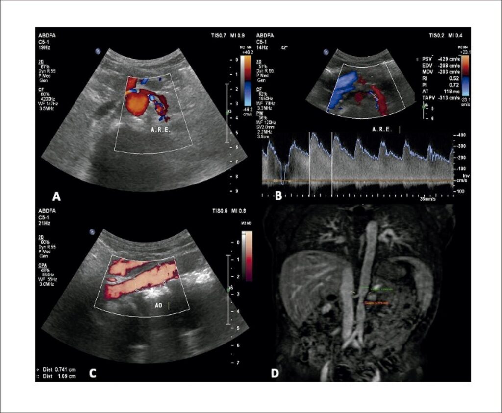
Takayasu arteritis (TA) is a rare large-vessel arteritis that primarily affects the aorta and its major branches. The greatest challenge is to identify disease activity, since therapeutic measures modify the clinical course of the disease.
Vascular ultrasonography (VUS) is a promising tool for characterizing vessel wall inflammation and monitoring hemodynamic changes in response to therapy.
[…]
The Value of Vascular Ultrasonography in Defining Inflammatory Activity in Takayasu Arteritis: Case Reports
BarrosFS, SantosSN, StorinoJ, FreireCMV, BarrosFS, DinisVG. The Value of Vascular Ultrasonography in Defining Inflammatory Activity in Takayasu Arteritis: Case Reports. Arq Bras Cardiol: Imagem cardiovasc. 2023;36(2):e20230021.
Barros,Fanilda Souto; Santos,Simone Nascimento dos; Storino,Joana; Freire,Cláudia Maria Vilas; Barros,Felipe Souto; Dinis,Valquiria Garcia. The Value of Vascular Ultrasonography in Defining Inflammatory Activity in Takayasu Arteritis: Case Reports. Arq Bras Cardiol: Imagem cardiovasc., v. 36, n. 2, e20230021, May. 2023.
Barros,F.S., Santos,S.N. , Storino,J., Freire,C.M.V., Barros,F.S., & Dinis,V.G. (2023). The Value of Vascular Ultrasonography in Defining Inflammatory Activity in Takayasu Arteritis: Case Reports. Arq Bras Cardiol: Imagem cardiovasc.,36(2), e20230021.
Barros,Fanilda Souto and Santos,Simone Nascimento dos and Storino,Joana and Freire,Cláudia Maria Vilas and Barros,Felipe Souto and Dinis,Valquiria Garcia. The Value of Vascular Ultrasonography in Defining Inflammatory Activity in Takayasu Arteritis: Case Reports. Arq Bras Cardiol: Imagem cardiovasc. [online]. 2023, vol. 36, n. 2, [cited 2026-03-06], e20230021. Available from: <https://www.abcimaging.org/article/the-value-of-vascular-ultrasonography-in-defining-inflammatory-activity-in-takayasu-arteritis-case-reports/>. ISSN .
Figure 1
– A) Color flow mapping shows lumen narrowing in the proximal course of the renal artery with flow turbulence; B) spectral Doppler analysis with PSV = 429cm/s and EDV = 208cm/s; C) measurement of the caliber of the abdominal aorta in the path with narrowing = 7.4mm, compared to the most distal path = 10.9mm; D) magnetic angioresonance of the abdominal aorta showing narrowing of the aorta in the path close to the origin of the mesenteric vessels.