ABC Imagem Cardiovasc. 2025; 38(4): e20250075
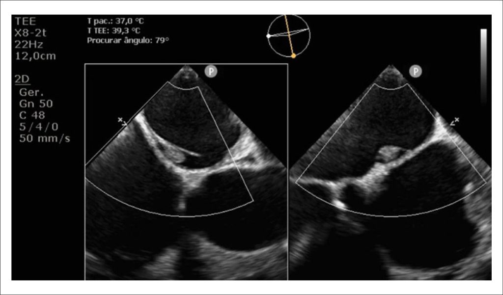
Left Atrial Septal Pouch Thrombosis Detected Before Electrical Cardioversion: a Rare Source of Embolism
DOI: 10.36660/abcimg.20250075i
A 59-year-old male patient with a history of systemic arterial hypertension was admitted to the emergency department with a seven-day history of chest pain and palpitations. On examination, he was hypotensive (93/70 mmHg) and tachycardic (150 bpm). A prominent systolic–diastolic murmur was heard along the right upper sternal border.
The initial investigation included electrocardiography (ECG) and Transthoracic Echocardiography (TTE). The ECG revealed atrial flutter, and the TTE showed biventricular dilation [Left Ventricular (LV) end-diastolic/end-systolic diameters: 60/51 mm; right ventricular (RV) basal diameter: 54 mm] and severe systolic dysfunction (LV ejection fraction: 19%; RV fractional area change: 16%). Additionally, a bicuspid aortic valve with mixed disease was identified: low-flow, low-gradient aortic stenosis (peak velocity: 3.1 m/s; peak/mean gradients: 39/23 mmHg; velocity ratio: 0.15; valve area: 0.7 cm, stroke volume index: 17 ml/m) and severe aortic regurgitation (pressure half-time: 170 ms; holodiastolic reversal in the descending aorta, end-diastolic velocity: 24 cm/s).
[…]
Keywords: Atrial Septum; Thrombosis; Transesophageal Echocardiography
80