Arq Bras Cardiol: Imagem cardiovasc. 2024; 37(3): e20240065
Fetal Intrapericardial Teratoma: Diagnostic and Therapeutic Challenges
DOI: 10.36660/abcimg.20240065i
Introduction
Primary cardiac tumors are rare in all age groups, with an incidence of 0.002% to 0.3% in autopsy series. During the prenatal period, cardiac tumors are highly uncommon and may be incidental findings in routine ultrasound assessment, which has become more frequent with the widespread use of imaging methods in the last decades. Fetal echocardiography is the main tool for detailed diagnosis of cardiac anatomy and function from the end of the first trimester until term., During fetal life, the most frequently found tumors are benign primary tumors, which include rhabdomyomas (the most common), as well as teratomas, fibromas, hemangiomas, and myxomas. The finding of malignant primary tumors (for example, rhabdomyosarcoma and fibrosarcoma) or metastatic tumors is exceptionally rare. In fetuses, cardiac tumors can be asymptomatic or lead to complications, such as arrhythmias, hydrops, obstruction of ventricular inflow or outflow, heart failure, and death.
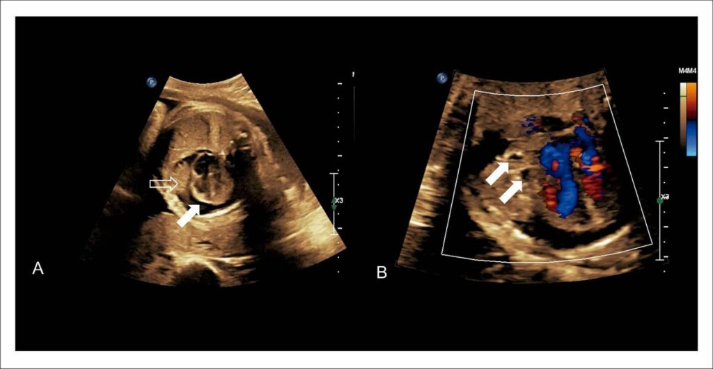
Teratomas, although they are usually benign, can have embryonic origin in one or more of the 3 germ layers, resulting in complex histology, with the possibility of simultaneous areas of mature and immature tissue. Intrapericardial teratomas are tumors with a low incidence, but they frequently invade the mediastinum and compress adjacent structures, leading to death. We report a case of intrapericardial teratoma diagnosed in a fetus at 22 weeks of gestation.
[…]
Keywords: Echocardiography; Pericardial Effusion; Prenatal Diagnosis
494