ABC Imagem Cardiovasc. 2025; 38(4): e20250067
Monitoring Myocardial Metabolic Changes in Lymphoma Patients Undergoing Chemotherapy Using FDG PET/CT
DOI: 10.36660/abcimg.20250067i
Abstract
Background:
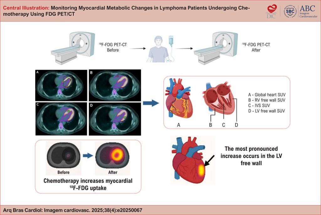
Cardiotoxicity is a serious adverse effect of chemotherapy, often identified only after irreversible myocardial damage has occurred. Positron emission tomography/computed tomography (PET/CT) using fluorine-18 fluorodeoxyglucose (18F-FDG) allows for the evaluation of myocardial glucose metabolism and may help detect early metabolic changes related to chemotherapy.
Objective:
To assess changes in 18F-FDG standardized uptake values (SUVs) across different cardiac regions before and after chemotherapy in patients with lymphoma, and to identify which region exhibits the greatest increase.
Methods:
This retrospective cohort study included 62 lymphoma patients who underwent 18F-FDG PET/CT before and after chemotherapy. SUV measurements were obtained in the left ventricular (LV) free wall, interventricular septum (IVS), right ventricular (RV) free wall, and global myocardium. Control regions included the liver and aorta. Pre- and post-treatment SUV values were compared to evaluate metabolic changes related to chemotherapy.
Results:
Myocardial 18F-FDG uptake increased significantly after chemotherapy across all cardiac regions, with the most pronounced rise observed in the LV free wall (maximum SUV increase of 73%, p<0.001). The RV free wall showed a non-significant increase in SUV, and no significant changes were observed in the liver or aorta.
Conclusions:
Chemotherapy was associated with a global increase in myocardial 18F-FDG uptake, with the most pronounced elevation observed in the LV free wall. This regional predominance highlights the LV free wall as the most sensitive site for detecting early metabolic changes potentially related to cardiotoxicity.
Keywords: Cardiotoxicity; Chemotherapy; Lymphoma; Myocardial metabolism
404