Arq Bras Cardiol: Imagem cardiovasc. 2024; 37(4): e20240102
Prevalence of Fetal Heart Disease in Pregnant Women Referred to a Specialized Service Due to Cardiac Abnormalities on Morphological Ultrasound
DOI: 10.36660/abcimg.20240102i
Abstract
Background:
Congenital Heart Disease (CHD) is a malformation of the heart and/or great vessels and is the third leading cause of neonatal death. It can be detected in utero by Morphological Ultrasound/Morphology Scan (MUS) and confirmed by Fetal Echocardiography (FE).
Objective:
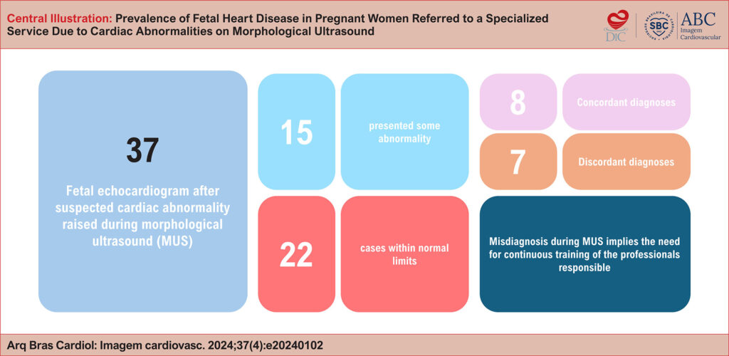
This study aims to determine whether heart disease identified by MUS in pregnant women at a maternal referral center in a southern Brazilian city is confirmed by FE.
Methods:
This observational, descriptive, and retrospective study was conducted from March 2019 to January 2023. It focused on pregnant women in the public health system, treated through the Unified Health System (SUS) at a maternal referral center in a southern Brazilian city, who had cardiac abnormalities detected on MUS and confirmed by FE.
Results:
Among the 37 pregnant women with suspected cardiac abnormalities on MUS, 15 cases were confirmed by FE, yielding a diagnostic agreement rate of 21.62% (n = 8) between the two exams. Ventricular Septal Defect (VSD) was the most prevalent heart defect, accounting for 20% of isolated cases. Of the 15 cases with cardiac alterations on FE, two were transferred to a specialized hospital and five died.
Conclusion:
Fetal cardiac abnormalities were confirmed in fewer than half of the women referred following MUS, with concordance between exams in only eight cases. Continuous training in MUS reduces costs and inconvenience. Professionals qualified in FE are essential for the early diagnosis of CHD, improving care, and reducing infant mortality.
473