Arq Bras Cardiol: Imagem cardiovasc 2023; 36(2): e20230055
My Approach to Three-Dimensional Echocardiography for Pathophysiological Classification of Tricuspid Regurgitation
DOI: 10.36660/abcimg.20230055i
Introduction
Although it was known as the “forgotten valve” in the past, there has been increasing interest in studying the tricuspid valve (TV) during the last two decades. Tricuspid regurgitation (TR) has been identified as a prognostic marker not only when associated with other cardiac diseases, like heart failure, mitral regurgitation, or aortic stenosis, – but also as an isolated entity. Understanding the pathophysiological mechanisms of this disease is of paramount importance, since new options of transcatheter devices and techniques are emerging, offering invasive treatment for high-risk patients unsuitable for surgical intervention.
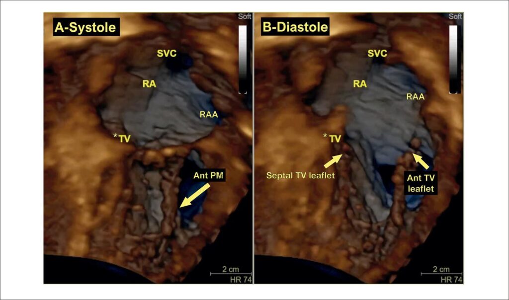
The TV is difficult to image in transesophageal echocardiography (TEE) for the following reasons: 1) the leaflets are much thinner compared to mitral leaflets, with greater anatomic variability; 2) it is an anterior structure, far away from the esophagus, with acoustic shadowing from the fibrous heart skeleton; and 3) it cannot be aligned to the esophageal probe in order to acquire en face views and requires use of lateral resolution. These restrictions may limit the ability of traditional TEE to evaluate the TV, making it necessary to complement the evaluation with special windows from the lower esophagus and transgastric views. Different from other cardiac valves, images of the TV obtained from transthoracic echocardiography (TTE) usually have better resolution than images obtained by TEE. Even though all protocols start with two-dimensional (2D) assessment of the TV, three-dimensional echocardiography (3DE) plays an important role in the evaluation of TV diseases, due not only to its ability to precisely depict the anatomy of the valve and the subvalvular apparatus, but also to its accuracy in quantitation of right ventricular (RV) and right atrial volumes and function and functional analysis of valvular dysfunction, especially for grading TR and evaluating dynamics of the tricuspid annulus (TA) through dedicated software. All of this information is of unparalleled importance for patient management and pre-procedural planning in surgical and transcatheter approaches.
[…]
2,209