Arq Bras Cardiol: Imagem cardiovasc. 2024; 37(2): e20240012
Use of a 3D Printer in the Prototyping of an Anatomical Model of the Human Heart with Morphophysiological Alterations
DOI: 10.36660/abcimg.20240012i
Abstract
Background:
Three-dimensional (3D) printing has a wide range of applications, including medicine and medical education. It presents itself as an alternative to traditional teaching-learning methods of human morphophysiology.
Objectives:
To report the initial experience in prototyping a 3D-printed anatomical model of the human heart with valvular heart disease, as well as its potential use in the active teaching-learning process in human morphophysiology.
Methods:
This is an experimental, descriptive study developed in three distinct stages: 1) Selection of the morphofunctional structure for manufacturing the 3D part; 2) design and adaptation of the object in 3D computer-aided design (CAD) software; and 3) printing and application of the prototype. Models extracted from the Thingiverse website, simulating cardiac structures, were used.
Results:
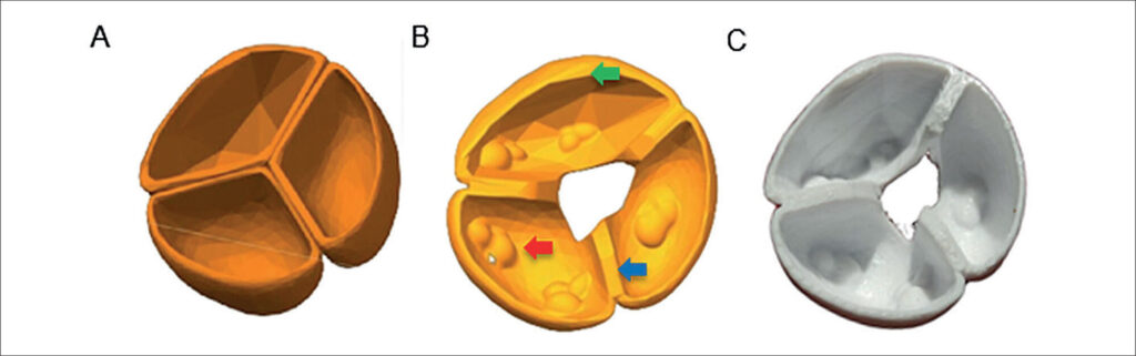
A cardiac prototype was 3D-printed to illustrate morphophysiological changes, including aortic stenosis and mitral insufficiency, modeled on the aortic and mitral valves, respectively.
Conclusion:
The valve diseases represented in the prototype accurately reflect the pathological changes used as the basis for the study, providing a detailed and precise view of the anatomy, which shows significant potential for enhancing the teaching-learning approaches in morphophysiology and medical pathology.
653