Arq Bras Cardiol: Imagem cardiovasc. 2024; 37(3): e20240044
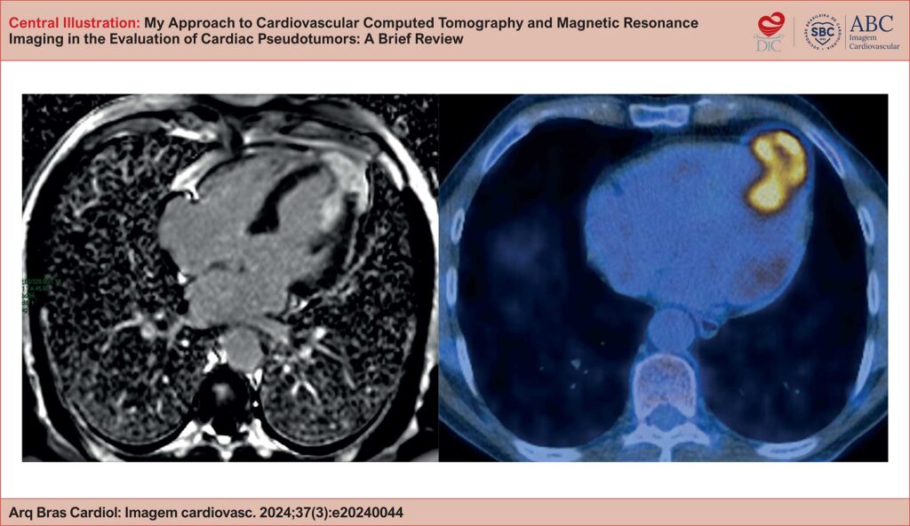
My Approach to Cardiovascular Computed Tomography and Magnetic Resonance Imaging in the Evaluation of Cardiac Pseudotumors: A Brief Review
DOI: 10.36660/abcimg.20240044i
Abstract
Cardiac pseudotumoral lesions are non-neoplastic conditions that are often overlooked in the differential diagnosis of cardiac masses. They present a variable clinical picture, ranging from asymptomatic to causing complications such as ventricular filling restriction and outflow tract obstruction. Echocardiography is the first-line imaging method but has limitations, such as dependence on the acoustic window and operator variability. However, a multimodality approach, including CT and MRI, is essential for seeking an accurate diagnosis. CT, with its excellent spatial resolution, allows for anatomical detailing, assessment of intralesional calcifications and fat, and contributes to therapeutic planning. MRI is preferred for tissue characterization and differentiation between benign and malignant lesions. Normal anatomical structures, such as the Eustachian valve and Chiari network, can be confused with thrombi or tumors, requiring correct identification. Thrombi are common in patients with atrial fibrillation or mitral valve disease, with MRI being important for differentiating them from neoplasms. Other pseudotumoral conditions include vegetation, gossypibomas, caseous calcification of the mitral valve annulus, and lipomatous hypertrophy of the interatrial septum. The integration of advanced cardiovascular imaging modalities is fundamental for the diagnosis and management of these lesions, optimizing patient care.
453