Arq Bras Cardiol: Imagem cardiovasc. 2021; 34(1): eabc131
Comparative Analysis of the Coronary Arteries Flow Pattern in Secondary Myocardial Hypertrophies and by Sarcomeric Mutation
DOI: 10.47593/2675-312X/20213401eabc131
Abstract
Background:
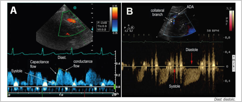
Coronary flow with a diastolic predominance increases two to five times in hyperemia, mediated by vasodilation (coronary flow reserve, CFR) and, in hypertrophy, relative ischemia may occur. In secondary hypertrophy (LVH), the flow, normal at rest, becomes ischemic due to increased demand. In hypertrophic cardiomyopathy (HCM) with perivascular fibrosis, collateral vessels appear to increase the irrigation of hypertrophied segments.
Objective:
To determine the coronary flow pattern in patients with secondary hypertrophy and hypertrophic cardiomyopathy, evaluating the coronary flow reserve.
Methods:
Coronary flow was evaluated in 34 patients with secondary hypertrophy, 24 with hypertrophic cardiomyopathy and in 16 controls. The anterior descending artery was detected with transthoracic Doppler with adequate equipment calibration. In the hypertrophic cardiomyopathy group, the flow of collaterals from the hypertrophic region was evaluated. In the control and secondary hypertrophy groups and in six patients in the hypertrophic cardiomyopathy group, the intravenous dipyridamole (0.84 mg) coronary flow reserve was calculated. The data were compared by variance with a significance of 5%.
Results:
In secondary hypertrophy there was an increase in mass index and blood pressure, and in hypertrophic cardiomyopathy an increase in relative thickness predominated. Ejection fraction and diastolic dysfunction were higher in the hypertrophic cardiomyopathy group. The coronary flow reserve was lower in the hypertrophic cardiomyopathy group, and flow of collaterals was also detected, with a reduction in the coronary flow reserve.
Conclusion:
the analysis of coronary circulation with transthoracic Doppler is possible in normal and hypertrophic individuals. Patients with secondary hypertrophy and hypertrophic cardiomyopathy have a decrease in the coronary flow reserve, and patients with hypertrophic cardiomyopathy show a hyper flow of dilated collateral vessels observed in the hypertrophic region, with a decrease in the coronary flow reserve.
149