Arq Bras Cardiol: Imagem cardiovasc. 2023; 36(4): e20230104
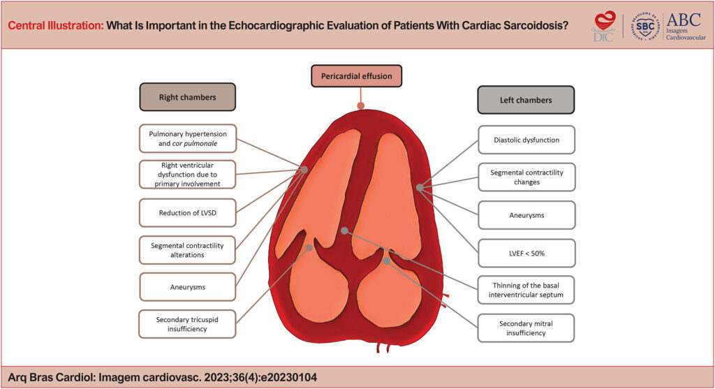
What Is Important in the Echocardiographic Evaluation of Patients With Cardiac Sarcoidosis?
DOI: 10.36660/abcimg.20230104i
Abstract
Sarcoidosis is a systemic condition, of unclear etiology, associated with the formation of non-necrotizing granulomas in several organs, and thoracic involvement in 90% of the cases. Cardiac impairment is detected in approximately 10% of the patients, reaching 25% in autopsy studies. It is in charge of about half the deaths in patients with sarcoidosis, and an important prognostic factor. Interventricular septum and the left ventricle free wall are the most affected regions, especially in the subepicardial portion. The development of changes in conduction (including atrioventricular block and ventricular arrhythmia) and heart failure are the most common manifestations. Diagnosis is challenging and often requires more advanced imaging examinations, such as positron emission tomography or late-enhancement cardiac magnetic resonance imaging. However, these examinations have high cost and are not so available. The conventional transthoracic echocardiography, on the other hand, is widely accessible, but presents later and little specific findings. The most important ones are the reduction of the left ventricle ejection fraction < 50% and the presence of abnormal tapering of the basal interventricular septum. Other segmental changes, especially when not correspondent to coronary territories, and aneurisms, are also relevant. Besides, there may be diastolic dysfunction, pericardial effusion and right ventricular dysfunction, both due to impairment primary or secondary to pulmonary hypertension. The most advanced ultrasound techniques, such as myocardial strain, myocardial work and elastography, are promising in the search of an earlier diagnosis at a lower cost.
Keywords: Sprains and Strains
1,318